3D-DOCTOR Related News
-
Multistation Becomes 3D-DOCTOR Reseller in France (July, 2012)
-
Twoplus Soft Becomes 3D-DOCTOR Reseller in Thailand (July, 2012)
-
Able Software Appoints AT Group Software as New Reseller in Mexico and Venezuela (June, 2012)
-
Able Software announces Widetech LTD as International Master Reseller (June, 2012)
-
3D-DOCTOR Is Used To Provide Medical Modeling Services (Aug. 2010)
-
Image Tilt Correction, 3D Angle Measurement (May 2008)
-
3D-DOCTOR Added New Region-Growing Based Segmentation (Nov. 2007)
-
Able Software Becomes SolidWorks Solution Partner (Oct. 2006)
-
SINGAPORE’S Republic Polytechnic USES 3D-DOCTOR FOR TEACHING (8, 2006)
-
3D-DOCTOR CHOSEN BY SINGAPORE’S Temasek Engineering School (Jan. 2004)
-
3D-DOCTOR NEW VERSION: COLOR FUSION, FOCUS FUSION AND MORE...
-
3D-DOCTOR 3.0: REAL TIME 3D SURFACE AND VOLUME RENDERING ON PC
-
3D-DOCTOR Supports DXF, STL, VRML, 3DS and Raw Triangle Formats
-
3D-DOCTOR is featured as the cover story in the Oct., 1998 issue of Advanced Imaging magazine.
3D-DOCTOR Major New Release Announced
Lexington, MA, USA - August 2, 2012 Able Software LLC., a global leader in 3D medical imaging and modeling software, announced today a release of its US FDA 510k cleared 3D-DOCTOR software. This new release delivers much refined and simplified user interface and improved 3D rendering and modeling performance. 3D-DOCTOR™ supports both grayscale and color images stored in DICOM, TIFF, Interfile, GIF, JPEG, PNG, BMP, PGM, RAW or other image file formats. 3D-DOCTOR creates 3D surface models and volume rendering from 2D cross-section images in real time on your PC. You can export the surface models to STL (ASCII and Binary), DXF, IGES, 3DS, OBJ, VRML, PLY, XYZ and other formats for surgical planning, simulation, quantitative analysis and rapid prototyping applications. 3D-DOCTOR is cleared by the US FDA (US Food and Drug Administration 510K clearance) for medical imaging and 3D visualization applications. It has been named the Top 3D Imaging Software by Scientific Computing & Instrumentation Magazine in the Year 2002 and Year 2000 Annual Technology Leaders Issue. Highlights of the new release include:
- Refined and simplified user interface, with a total redesign of the tool buttons. The tool bars now use a large size with brighter graphics design so they can be located and used easily.
- Image and 3D rendering display windows no longer use the traditional scrollbars so more display space is used for image content. Image panning and scrolling is now done easily with the mouse.
- Reorganization of the user menu items. Related functions are grouped together and in the order they are used to finish a certain task.
- Improved international support, for the Japanese and Chinese user interface. Support to other languages are being added.
- The online help has been improved significantly with added tutorials and links to tutorial video clips.
- Performance improvements are implemented to increase the speed of 3D surface rendering and mesh model generation.
Multistation Becomes 3D-DOCTOR Reseller in France
Lexington, MA, USA - July 13, 2012 Able Software LLC., a global leader in 3D medical imaging and modeling software and the developer of the US FDA 510k cleared 3D-DOCTOR software, announced today a reseller partnership with Multistation SAS (www.multistation.com), with offices in France and North Africa. Since 1987, Multistation SAS delivers worldwide industrial equipments of first quality for different applications like automotive, aerospace, railways, energy, defence, marine, education and medical… but also in a certain number of very diversified niches like the jewelry or complete solution for dental applications. Multistation SAS will resell the 3D-DOCTOR software in France. Multistation SAS will facilitate the introduction of the 3D-DOCTOR™ software solutions to customers, providing an advanced 3D modeling, image processing, and measurement software for MRI, CT, PET, microscopy, scientific, and industrial 3D imaging applications. Multistation SAS is assigned as an authorized reseller in the territory by our International master reseller Widetech Ltd and will be reporting directly to Widetech (http://www.widetech.co.il). 3D-DOCTOR™ supports both grayscale and color images stored in DICOM, TIFF, Interfile, GIF, JPEG, PNG, BMP, PGM, RAW or other image file formats. 3D-DOCTOR creates 3D surface models and volume rendering from 2D cross-section images in real time on your PC. You can export the surface models to STL (ASCII and Binary), DXF, IGES, 3DS, OBJ, VRML, PLY, XYZ and other formats for surgical planning, simulation, quantitative analysis and rapid prototyping applications. 3D-DOCTOR is cleared by the US FDA (US Food and Drug Administration 510K clearance) for medical imaging and 3D visualization applications. It has been named the Top 3D Imaging Software by Scientific Computing & Instrumentation Magazine in the Year 2002 and Year 2000 Annual Technology Leaders Issue. "We are very excited to have Multistation SAS as a reseller to offer our 3D-DOCTOR to their customers throughout France," said Dr. Ted Wu, Founder and CEO of Able Software. "Their in-depth technical knowledge in 3D modeling and 3D rapid prototyping applications will enable them to leverage the power of 3D-DOCTOR to offer their customers better and cost-effective solutions." "3D-DOCTOR is a genius software which will help us to provide complete solutions to our educational, medical, industrial and design customers; we will increase our capacity of value added reselling in 3D digital , medical and additive manufacturing ", added Mr. Yannick Loisance, Founder and CEO of Multistation.r.a.p.s AB Resells 3D-DOCTOR in Sweden
Lexington, MA, USA - July 12, 2012 Able Software LLC., a global leader in 3D medical imaging and modeling software and the developer of the US FDA 510k cleared 3D-DOCTOR software, announced today a reseller partnership with r.a.p.s AB, with offices in Sweden. Known for its expertise and high level of service, r.a.p.s AB (http://raps.se) is a distributor of 3D imaging and CAD software solutions and 3D Printers. r.a.p.s AB will resell the 3D-DOCTOR software in Sweden. r.a.p.s AB will facilitate the introduction of the 3D-DOCTOR™ software solutions to customers, providing an advanced 3D modeling, image processing, and measurement software for MRI, CT, PET, microscopy, scientific, and industrial 3D imaging applications. r.a.p.s AB is assigned as an authorized reseller in the territory by our International master reseller Widetech Ltd and will be reporting directly to Widetech (http://www.widetech.co.il).3D-DOCTOR™ supports both grayscale and color images stored in DICOM, TIFF, Interfile, GIF, JPEG, PNG, BMP, PGM, RAW or other image file formats. 3D-DOCTOR creates 3D surface models and volume rendering from 2D cross-section images in real time on your PC. You can export the surface models to STL (ASCII and Binary), DXF, IGES, 3DS, OBJ, VRML, PLY, XYZ and other formats for surgical planning, simulation, quantitative analysis and rapid prototyping applications. 3D-DOCTOR is cleared by the US FDA (US Food and Drug Administration 510K clearance) for medical imaging and 3D visualization applications. It has been named the Top 3D Imaging Software by Scientific Computing & Instrumentation Magazine in the Year 2002 and Year 2000 Annual Technology Leaders Issue.
Korimo Becomes 3D-DOCTOR Reseller in Slovakia
Lexington, MA, USA - July 10, 2012 Able Software LLC., a global leader in 3D medical imaging and modeling software and the developer of the US FDA 510k cleared 3D-DOCTOR software, announced today a reseller partnership with Korimo s.r.o., with offices in Slovakia. Known for its expertise and high level of service, Korimo s.r.o. (http://www.korimo.sk/) is a distributor of 3D imaging and CAD software solutions and 3D Printers. Korimo s.r.o. will resell the 3D-DOCTOR software in Slovakia. Korimo s.r.o. will facilitate the introduction of the 3D-DOCTOR™ software solutions to customers, providing an advanced 3D modeling, image processing, and measurement software for MRI, CT, PET, microscopy, scientific, and industrial 3D imaging applications. Korimo s.r.o. is assigned as an authorized reseller in the territory by our International master reseller Widetech Ltd and will be reporting directly to Widetech (http://www.widetech.co.il). 3D-DOCTOR™ supports both grayscale and color images stored in DICOM, TIFF, Interfile, GIF, JPEG, PNG, BMP, PGM, RAW or other image file formats. 3D-DOCTOR creates 3D surface models and volume rendering from 2D cross-section images in real time on your PC. You can export the surface models to STL (ASCII and Binary), DXF, IGES, 3DS, OBJ, VRML, PLY, XYZ and other formats for surgical planning, simulation, quantitative analysis and rapid prototyping applications. 3D-DOCTOR is cleared by the US FDA (US Food and Drug Administration 510K clearance) for medical imaging and 3D visualization applications. It has been named the Top 3D Imaging Software by Scientific Computing & Instrumentation Magazine in the Year 2002 and Year 2000 Annual Technology Leaders Issue. “We are very excited to have Korimo s.r.o. as a reseller to offer our 3D-DOCTOR to their customers throughout Slovakia,” said Dr. Ted Wu, Founder and CEO of Able Software. “Their in-depth technical knowledge in 3D modeling and 3D rapid prototyping applications will enable them to leverage the power of 3D-DOCTOR to offer their customers better and cost-effective solutions.”Twoplus Soft Becomes 3D-DOCTOR Reseller in Thailand
Lexington, MA, USA - July 5, 2012 Able Software LLC., a global leader in 3D medical imaging and modeling software and the developer of the US FDA 510k cleared 3D-DOCTOR software, announced today a reseller partnership with Twoplus Soft Co., with offices in Thailand. Known for its expertise and high level of service, Twoplus Soft Co. (www.twoplusssoft.com) is a distributor of 3D imaging and CAD software solutions and 3D Printers for the Healthcare, Architecture, Engineering and Construction. Twoplus Soft Co. will resell the 3D-DOCTOR software in Thailand. Twoplus Soft Co. will facilitate the introduction of the 3D-DOCTOR™ software solutions to customers, providing an advanced 3D modeling, image processing, and measurement software for MRI, CT, PET, microscopy, scientific, and industrial 3D imaging applications. 3D-DOCTOR™ supports both grayscale and color images stored in DICOM, TIFF, Interfile, GIF, JPEG, PNG, BMP, PGM, RAW or other image file formats. 3D-DOCTOR creates 3D surface models and volume rendering from 2D cross-section images in real time on your PC. You can export the surface models to STL (ASCII and Binary), DXF, IGES, 3DS, OBJ, VRML, PLY, XYZ and other formats for surgical planning, simulation, quantitative analysis and rapid prototyping applications. 3D-DOCTOR is cleared by the US FDA (US Food and Drug Administration 510K clearance) for medical imaging and 3D visualization applications. It has been named the Top 3D Imaging Software by Scientific Computing & Instrumentation Magazine in the Year 2002 and Year 2000 Annual Technology Leaders Issue.Able Software Adds new Reseller in Romania
Lexington, MA, USA - July 2, 2012 Able Software LLC., a global leader in 3D medical imaging and modeling software and the developer of the US FDA 510k cleared 3D-DOCTOR software, announced today a reseller partnership with Logicad Solutions srl located in Romania. Known for its expertise and high level of service in the CAD/CAM market, Logicad Solutions (http://www.logicad.ro) is a distributor of 3D imaging and CAD software solutions and 3D Printers. Logicad Solutions will resell the 3D-DOCTOR software in Mexico and Venezuela. Logicad Solutions will facilitate the introduction of the 3D-DOCTOR™ software solutions to customers, providing an advanced 3D modeling, image processing, and measurement software for MRI, CT, PET, microscopy, scientific, and industrial 3D imaging applications. Logicad Solutions is assigned as an authorized reseller in the territory by our International master reseller Widetech Ltd and will be reporting directly to Widetech (http://www.widetech.co.il). 3D-DOCTOR™ supports both grayscale and color images stored in DICOM, TIFF, Interfile, GIF, JPEG, PNG, BMP, PGM, RAW or other image file formats. 3D-DOCTOR creates 3D surface models and volume rendering from 2D cross-section images in real time on your PC. You can export the surface models to STL (ASCII and Binary), DXF, IGES, 3DS, OBJ, VRML, PLY, XYZ and other formats for surgical planning, simulation, quantitative analysis and rapid prototyping applications. 3D-DOCTOR is cleared by the US FDA (US Food and Drug Administration 510K clearance) for medical imaging and 3D visualization applications. It has been named the Top 3D Imaging Software by Scientific Computing & Instrumentation Magazine in the Year 2002 and Year 2000 Annual Technology Leaders Issue.Able Software Announces new Reseller in Mexico and Venezuela
Lexington, MA, USA - June 27, 2012 Able Software LLC., a global leader in 3D medical imaging and modeling software and the developer of the US FDA 510k cleared 3D-DOCTOR software, announced today a reseller partnership with AT Group Software, with offices in Mexico and Venezuela. Known for its expertise and high level of service, AT Group Software (http://www.atgroup.com.mx/) is a distributor of 3D imaging and CAD software solutions and 3D Printers. AT Group Software will resell the 3D-DOCTOR software in Mexico and Venezuela. AT Group will facilitate the introduction of the 3D-DOCTOR™ software solutions to customers, providing an advanced 3D modeling, image processing, and measurement software for MRI, CT, PET, microscopy, scientific, and industrial 3D imaging applications. AT Group is assigned as an authorized reseller in the territory by our International master reseller Widetech Ltd and will be reporting directly to Widetech (http://www.widetech.co.il).3D-DOCTOR™ supports both grayscale and color images stored in DICOM, TIFF, Interfile, GIF, JPEG, PNG, BMP, PGM, RAW or other image file formats. 3D-DOCTOR creates 3D surface models and volume rendering from 2D cross-section images in real time on your PC. You can export the surface models to STL (ASCII and Binary), DXF, IGES, 3DS, OBJ, VRML, PLY, XYZ and other formats for surgical planning, simulation, quantitative analysis and rapid prototyping applications. 3D-DOCTOR is cleared by the US FDA (US Food and Drug Administration 510K clearance) for medical imaging and 3D visualization applications. It has been named the Top 3D Imaging Software by Scientific Computing & Instrumentation Magazine in the Year 2002 and Year 2000 Annual Technology Leaders Issue.
"We are very excited to have AT Group Software as a reseller to offer our 3D-DOCTOR to their customers throughout Mexico and Venezuela," said Dr. Ted Wu, Founder and CEO of Able Software. "Their in-depth technical knowledge in 3D modeling and 3D rapid prototyping applications will enable them to leverage the power of 3D-DOCTOR to offer their customers better and cost-effective solutions."
ABLE Software Announces Widetech LTD as International Master Reseller
LEXINGTON, MA U.S.A - June16, 2012 – ABLE Software™ Corporation, a leading supplier of 3D-DOCTOR™ Software for medical image processing and modeling, today announced the selection of Widetech LTD as its International Master Reseller.
ABLE Software ™ Corporation has entered into a multi-year agreement
with Widetech LTD to develop and manage a growing Authorized Reseller
network in the International markets. Through an expanded base of
resellers, Widetech LTD will facilitate the introduction of the
3D-DOCTOR™ software solutions to customers, providing an advanced
3D modeling, image processing, and measurement software for MRI,
CT, PET, microscopy, scientific, and industrial 3D imaging
applications.
3D-DOCTOR™ supports both grayscale and color images stored in DICOM,
TIFF, Interfile, GIF, JPEG, PNG, BMP, PGM, RAW or other image file
formats. 3D-DOCTOR creates 3D surface models and volume rendering from
2D cross-section images in real time on your PC. You can export the
surface models to STL (ASCII and Binary), DXF, IGES, 3DS, OBJ, VRML,
PLY, XYZ and other formats for surgical planning, simulation,
quantitative analysis and rapid prototyping applications.
3D-DOCTOR is approved by the US FDA (US Food and Drug Administration
510K clearance) for medical imaging and 3D visualization applications.
It has been named the Top 3D Imaging Software by Scientific Computing
& Instrumentation Magazine in the Year 2002 and Year 2000 Annual
Technology Leaders Issue.
“We are very excited about this cooperation with ABLE Software”,
said Eldad Sayada, CEO of Widetech LTD, “We are committed to delivering
simple solution for educational institutions, research groups and
hospitals. The ability for use this software with user on a
simple PC changed the availability for processing imaging created by CT
and MRI”. Furthermore, Mr. Sayada added it will open up endless
possibilities for researchers to use off the shelf product for their
research”.
Ted Wu, PhD, Founder and CEO of ABLE Software, indicated that with the
appointment of Widetech LTD as its International Master Reseller, the
sales channels will expand rapidly in the worldwide medical imaging and
3D modeling market. Added Dr. Wu, “Widetech LTD’s years of sales
experience and expertise in medical imaging and modeling applications
will help us to reach more users and establish a more complete reseller
network for different market segments. We’ll work closely with Widetech
LTD to support and serve the customers and develop the next generation
products.”
About ABLE Software
Able Software LLC. is a leading imaging software developer with its
headquarter in the US since 1994. Able Software has 2 main
software products, 3D-DOCTOR and R2V.
The 3D-DOCTOR software is a US FDA 510K cleared medical imaging, 3D
modeling and visualization software. It's currently being used by
leading hospitals, medical schools and surgeons around the world.
The R2V software is currently being used in more than 100 countries for
map digitizing, CAD drawing conversion and geographic information
system (GIS) applications.
About Widetech LTD
Widetech LTD (http://www.widetech.co.il) is a professional
international distribution organization for a range of products in 3D
engineering tools and product support. Widetech was established in 2004
and since then Eldad Sayada is leading the company into the
international market. Widetech professional marketing services range
from 3D printers, 3D scanners, CMM inspection and QA measurements and
CAD related products.
ABLE Software Press Contact
Ted Wu, PhD
Founder and CEO, ABLE Software Corporation
ywu@ablesw.com
Widetech Press Contact
Eldad Sayada
CEO, Widetech LTD
eldad@widetech.co.il
+(972)-54-80.81.810
(Aug. 31, 2010 Boxborough, MA) Voxelor Inc., a startup company founded by a group of veteran medical imaging professionals, has been officially launched in Boxborough, MA to provide 3D medical modeling services to doctors, surgeons and medical researchers.
The company creates both digital and physical 3D models from patient CT or MRI scans according to the doctor's prescription. The models are normally used for diagnosis, treatment, surgical planning, patient education and other clinical applications.
Voxelor Inc. has partnered with industry leaders, including Able Software LLC. (http://www.ablesw.com) and 3D Systems (http://www.3dsystems.com), to gain access to the latest technologies in medical modeling software and rapid prototyping.
Able Software's 3D-DOCTOR software is a leading 3D medical imaging and modeling software. It has received FDA 510K clearance and is currently being used by hospitals and medical schools around the world.
3D Systems is a leading provider of 3-D Printing, Rapid Prototyping and Manufacturing systems and parts solutions. 3D Systems' technologies are being used today for medical, dental, industrial manufacturing and rapid prototyping applications.
"Our goal is to provide high quality virtual and tactile medical models to doctors and surgeons for better patient care. Our website, www.voxelor.com, was designed to provide a clear and simple to use interface for doctors to upload their data and get the models created. No training is needed to use the website. Although the site is fairly new, many doctors have already expressed interest in using our services.", said Dr. Chen Yi, President and COO of Voxelor Inc.
Voxelor is
headquartered in Boxborough, MA. It will provide 3D medical modeling
services worldwide. Regional offices providing sales and technical
support are being set up in other countries.
Image Tilt Correction, 3D Angle Measurement
(5/20/2008, LEXINGTON, MA) – Able Software LLC. has released a new version of 3D-DOCTOR software. This version implemented gantry tilt correction, as well as 3D angle measurement functions.
This new version includes the following new functions and improvements:
A new "Add Folder" option is added to the “New Stack” command. This function will search all files and subfolders within the selected folder for image files and add them to a stack list. It solves the problem where some CT scanners save DICOM files in many subfolders.
A new 3D angle measurement tool is added to the surface rendering window. Select “Tool/Measure Angle” to start the tool. Click 3 points on a model to get the angle measured.
New “Image/Tilt Correction” function is implemented to correct gantry tilt image distortion. Some CT image slices (most are in DICOM format) are acquired with a tilted gantry so the slices are not perfectly perpendicular to the main axis. This new function will use the angle stored in the DICOM header to compensate the tilt and generate image slices that are perpendicular to the main axis for segmentation and 3D rendering applications.
Added “Line Width” attribute for the object boundary display. To change the boundary line display width, use the “Edit/Object Settings” command and then click on the “Line Type” button.
Enabled editing options for the regions of interest (ROI) created from object boundaries (using the “Edit/ROI/ROI from Boundaries command). ROIs on different image planes can be edited and deleted using the ROI editor.
Added the “Move Note” function to the “Annotation Editor” to move an existing annotation to a different location.
The “Delete Node” tool under the “Boundary Editor” has been improved to allow removing nodes continuously. When the “Delete Node” tool is selected, pressing down the left mouse button will change the tool to a node eraser and boundary nodes touched by the eraser will be removed.
Improved the "Copy Boundary" tool under the “Boundary Editor”. One can draw a selection rectangle to select a group of boundaries and then use the “Control-C” key to copy. The boundaries can be pasted to a different image plane using the “Paste” or “Control-V” key.
Additional image output file formats are added to the “File/Save Window” command. The window image can now be saved to BMP, TIFF, JPEG, DICOM and other formats.
The 3D “Volume and Surface Area Calculation” command now uses an area-weighted algorithm to calculate the centroid of a 3D object.
HTML based online help is now supported in 3D-DOCTOR.
New image conversion function to convert 1-bit image to 8-bit grayscale.
3D-DOCTOR Added New Region-Growing Based Segmentation
(11/1/2007, LEXINGTON, MA) – Able Software Corp. has released a new version of 3D-DOCTOR software. This version implemented a new region-growing based segmentation that is more effective separating soft tissues in a CT or MRI image.
Other improvements include image reslicing functions, quantitative analysis and 3D measurement functions.
Able
Software’s 3D-DOCTOR is a FDA
(United States Food and Drug Administration) approved 3D medical
imaging and modeling software. It is currently being used by medical
schools, hospitals and research organizations around the world.
3D-DOCTOR creates 3D models from CT/MRI scans for 3D medical modeling,
diagnosis, surgical planning and quantitative analysis application. 3D
models can be exported to SolidWorks for editing and analysis.
3D-DOCTOR supports many commonly used graphics formats, including STL,
VRML, IGES, 3DS, DXF, OBJ, PLY and others for rapid prototyping and 3D
visualization applications.
Headquartered in LEXINGTON, MA, ABLE SOFTWARE CORP. is a leading developer in Windows-based 3D imaging and raster to vector conversion software market, with users in more than 100 countries. ABLE SOFTWARE was founded in 1993.
Free 3D-DOCTOR Viewer is Available for Download
(May 2007 Lexington, MA) - Able Software LLC. has released a new 3D-DOCTOR Viewer for visualizing 3D rendering and image files.
The
Viewer is designed as a simple display tool for users who do
not have 3D-DOCTOR software. When a complex 3D rendering is created
using 3D-DOCTOR's rendering functions,
the 3D file can be distributed together with the Viewer so others can
see the
full 3D display with all the controls, such as rotation, zoom in and
out and
animation.
To show a 3D rendering to others, simply save the volume rendering to a
volume (.vol) file or surface rendering to a surface file (.suf) and
send the files with the 3D-DOCTOR viewer. The Viewer will display the
3D rendering exactly the same
way as using 3D-DOCTOR itself. With the Viewer, a 3D display can be
rotated, adjusted and animated.
The Viewer is FREE for download.
More info can be found at: http://www.ablesw.com/3d-doctor/3dviewer.html
Able Software Becomes SolidWorks Solution Partner
(10/16/2006
LEXINGTON, MA) – Able Software LLC. has
become a Solution Partner with SolidWorks Corporation.
Able Software is the developer of 3D-DOCTOR and R2V
software products. SolidWorks develops 3D mechanical design software.
Able
Software’s 3D-DOCTOR is a FDA
(United States Food and Drug Administration) approved 3D medical
imaging and modeling software. It is currently being used by medical
schools, hospitals and research organizations around the world.
3D-DOCTOR creates 3D models from CT/MRI scans for 3D medical modeling,
diagnosis, surgical planning and quantitative analysis application. 3D
models can be exported to SolidWorks for editing and analysis.
3D-DOCTOR supports many commonly used graphics formats, including STL,
VRML, IGES, 3DS, DXF, OBJ, PLY and others for rapid prototyping and 3D
visualization applications.
Able
Software’s R2V software is an automated raster to vector conversion
software. It converts scanned maps and drawings into a vector format so
the data can be used in a design or mapping software.
SolidWorks
Corporation was the first company to develop powerful 3D mechanical
design software. SolidWorks®
software is the world’s #1 3D design software for the mainstream market
based on the number of users in production and sales.
3D-DOCTOR New Version 4.0
(Sept. 2006, Lexington, MA, USA) - Able Software has released a new version 4.0 of 3D-DOCTOR, the vector-based 3D imaging, modeling and measurement software for CT, MRI, microscopy and volumetric images.
This new version has added import function to load 32-bit image files, raw image with signed pixels and raw image in ASCII text format.
An interactive 3D image registration function is implemented to register images of different modalities, such as CT, MRI and PET. The registration function displays both the source and the target image in 3D and interactively adjusts (rotate, move and stretch) the source image until it fits the target the image. A control point based registration function is also available for multi-modality 3D image registration and fusion.
The 3D surface modeling algorithm has been enhanced to provide faster rendering and generate high quality 3D mesh models from CT/MRI scans. 3D models are used for 3D measurement, volume calculation, surgical and treatment planning, surgical simulation, and 3D rapid prototyping applications.
A new animation function is implemented to create movies and 3D simulations from 3D rendering. The tissue display properties can set as transparent, opaque, wire frame or with texture map during the animation. Objects can be moved, scaled, hidden or made visible in the animation process.
The improved volume rendering function creates real-time 3D image visualization. It supports both color and grayscale rendering using either opaque or transparent voxels. The volume rendering uses either the entire image volume, a portion defined by regions of interest (ROI), or a portion defined by the user interactively. Tissues with different density can be included or excluded in the rendering by changing their opacity property.
3D-DOCTOR is developed by Able Software Corp., a 3D imaging software developer with users in more than 100 countries. 3D-DOCTOR has been approved by US FDA (United States Food and Drug Administration) for medical imaging applications and is currently being used by research organizations and hospitals around the world. 3D-DOCTOR creates 3D computer models from medical images stored in DICOM, TIFF, Interfile, BMP, JPEG, PNG, GIF and raw formats. Both color and grayscale images are supported. The generated 3D models can be exported to many commonly used graphics formats, including VRML, IGES, 3DS, DXF, OBJ, STL, PLY and others for 3D visualization and modeling applications.
SINGAPORE’S Republic Polytechnic USES 3D-DOCTOR FOR TEACHING
(8/16/2006 LEXINGTON, MA) – Singapore’s Republic Polytechnic (www.rp.sg) has purchased 55 licenses of 3D-DOCTOR software to equip the department of Biomedical Electronics. 3D-DOCTOR will be used to teach the subject of “Fundamental Concepts of Medical Imaging and Processing” and for 3D modeling and rapid prototyping applications.
Republic Polytechnic is a leading educational institution in Singapore with its well-known emphasis on the use of technology to support learning. The Biomedical Electronics program provides students with the skills and practical knowledge in the areas of sensors and instrument systems in biomedicine, imaging techniques and signal processing. The program focuses on the integrated use of electronics, electrical and computer technologies in medical devices used in the biomedical industry to day.
3D-DOCTOR is developed by Able Software Corp., a 3D imaging software developer with users in more than 100 countries. 3D-DOCTOR has been approved by US FDA (United States Food and Drug Administration) for medical imaging applications. It is currently being used by research organizations and hospitals around the world. 3D-DOCTOR creates 3D models from CT/MRI scans for 3D medical modeling, diagnosis, surgical planning and quantitative analysis application. 3D models can be exported to many commonly used graphics formats, including STL, VRML, IGES, 3DS, DXF, OBJ and others for rapid prototyping and 3D visualization applications.
3D-DOCTOR NEW RELEASE: ENHANCED 3D MEDICAL MODELING FROM CT/MRI
(5/3/2005 LEXINGTON, MA) – ABLE SOFTWARE released a new version of 3D-DOCTOR, the vector-based 3D imaging, modeling and measurement software for CT, MRI, microscopy and volumetric images.
The 3D surface modeling algorithm has been enhanced to provide faster rendering and high quality models. Polygon-based mesh models are created from CT/MRI images in DICOM or other image formats. 3D models can be used for 3D measurement and volume calculation, surgical and treatment planning, surgical simulation, and 3D rapid prototyping applications.
The new “smooth shading” volume rendering function can be used for real-time 3D image visualization. It supports both color and grayscale rendering using either opaque or transparent voxels. The volume rendering uses either the entire image volume, a portion defined by regions of interest (ROI), or a portion defined by object boundaries.
New boundary tracing functions have been implemented for quick and easy object boundary definition using a touch screen or a tablet. Object boundaries are used to create 3D mesh models and generate quantitative measurement reports.
3D-DOCTOR is developed by Able Software LLC., a 3D imaging software developer with users in more than 100 countries. 3D-DOCTOR has been approved by US FDA (United States Food and Drug Administration) for medical imaging applications and is currently being used by research organizations and hospitals around the world. 3D-DOCTOR creates 3D computer models from medical images stored in DICOM, TIFF, Interfile, BMP, JPEG, PNG, GIF and raw formats. Both color and grayscale images are supported. The generated 3D models can be exported to many commonly used graphics formats, including VRML, IGES, 3DS, DXF, OBJ, STL and others for 3D visualization and modeling applications.
3D-DOCTOR CHOSEN BY SINGAPORE’S Temasek Engineering School
(1/22/2004 LEXINGTON, MA) – Singapore’s Temasek Engineering School recently purchased a multiple-node site license of 3D-DOCTOR software to equip their Rapid Prototyping Technology Unit for 3D modeling and rapid prototyping applications.
Temasek Engineering School is a leading university in Singapore for advanced engineering research and education. The licenses of 3D-DOCTOR are installed at the Rapid Prototyping Technology (RPT) Unit for creating 3D computer and physical models from medical and industrial images. The academic mission of the RPT unit is to educate and create awareness of Rapid Prototyping Technology in the industry, and to improve and enhance the rapid prototyping technology through research and industrial projects. The RPT unit is focused on 3D modeling, rapid prototyping, rapid tooling, laser scanning, sheet metal forming and injection molding for medical, industrial and scientific applications.
3D-DOCTOR is developed by Able Software LLC. a 3D imaging software developer with users in more than 100 countries. 3D-DOCTOR has been approved by US FDA (United States Food and Drug Administration) for medical imaging applications and is currently being used by research organizations and hospitals around the world. 3D-DOCTOR creates 3D computer models from medical images stored in DICOM, TIFF, Interfile, BMP, JPEG, PNG, GIF and raw formats. Both color and grayscale images are supported. The generated 3D models can be exported to many commonly used graphics formats, including VRML, IGES, 3DS, DXF, OBJ, STL and others for 3D visualization and modeling applications.
3D-DOCTOR ADDS ADVANCED ANIMATION AND MODELING
(2/12/2003
LEXINGTON, MA) -- ABLE SOFTWARE announced a new update of
3D-DOCTOR version 3.5, a 3D image visualization, rendering and
measurement
software for CT, MRI, microscopy and scientific images.
This new release has implemented advanced Animation Control functions to create 3D simulation and animation of multiple objects for surgical planning and modeling applications. With the new functions, a user can define the animation path of each 3D object with more than 9 degrees of freedom, including the size, location, orientation and the viewing angle. The animation can be saved to a digital movie file for remote viewing and presentation. Other implementations include functions to adjust the size, location and orientation of 3D models, as well as integrated display of raster image and polygon-based 3D models. 3D models created from different sources can be combined into a single coordinate system for display and comparison analysis.
3D-DOCTOR supports image files stored in DICOM, TIFF, Interfile, BMP, JPEG, PNG and raw formats. 3D surface models are created from cross-section image slices and saved to a 3D format, including VRML, IGES, 3DS, DXF, OBJ, STL and other formats for visualization and surgical modeling and planning applications. The 3D image registration function registers multi-modality images for comparison and image fusion applications. 3D-DOCTOR is FDA 510K Cleared for medical imaging applications and currently being used by leading research organizations and hospitals around the world.
3D-DOCTOR IS NAMED AGAIN THE TOP 3D Imaging Software by Scientific Computing & Instrumentation Magazine FOR the Year 2002
3D-DOCTOR 3.5 is 3-D image visualization, rendering and measurement software for CT, MRI, microscopy and scientific images. It includes functions for quantitative analysis and 3-D color image processing and real time 3-D surface and volume rendering algorithms. The software supports image files stored in DICOM, TIFF Interfile, BMP, JPEG, PNG and raw formats. 3-D surface models are created from cross-section image slices and saved to 3-D formats, including VRML, IGES, 3DS, DXF, OBJ, and STL.
3D-DOCTOR NEW VERSION 3.5
(4/11/2002
LEXINGTON, MA) -- ABLE SOFTWARE announced a new release of
3D-DOCTOR version 3.5, a 3D image visualization, rendering and
measurement
software for CT, MRI, microscopy and scientific images.
This new release includes new functions for quantitative analysis and 3D color image processing and improvements to the real time 3D surface and volume rendering algorithms.
The
quantitative analysis functions are used to calculate histogram,
size and volume for tumor size measurement and cancer
treatment applications. 3D image processing functions, such as
color image segmentation, image registration and fusion have
been implemented. 3D-DOCTOR supports image files
stored in DICOM, TIFF, Interfile, BMP, JPEG, PNG and raw formats.
3D surface models are created from cross-section image slices
and saved to a 3D
format, including VRML, IGES, 3DS, DXF, OBJ, STL and other formats
for visualization and surgical modeling and planning
applications. The 3D image registration function registers
multi-modality images for comparison and image fusion
applications.
3D-DOCTOR is FDA 510K Cleared for medical imaging applications and
currently being used by leading research organizations and
hospitals around the world, including Mass. General Hospital,
Harvard Medical School, MIT, Brigham Women Hospital, Stanford
Medical School, MD Anderson Cancer Center,
Memorial Sloan-Kettering Cancer Center, SUNY Buffalo Imaging
Center, Univ. of Minnesota Imaging Center, National Hospital
of Norway, VA Medical Center, US
Army Surgical Research Institute and many others.
3D-DOCTOR RECEIVES FDA CLEARANCE
(March 14, 2001) Able Software today announced that the US FDA (US Food and Drug Administration) has granted the company 510(k) clearance to market its 3D-DOCTOR software for medical imaging applications. 3D-DOCTOR is an advanced 3D imaging, rendering, and measurement software for CT, MRI, microscopy, and other volumetric images.
3D-DOCTOR software visualizes image data from CT, MRI, and microscopy imaging devices in DICOM, TIFF, JPEG, BMP, and other vendor specific file formats. The proprietary vector-based surface rendering algorithm creates 3D surface models from cross-section images in real time, on a low-cost standard PC. The 3D surface models created with this algorithm result in much less surface triangles than raster-based methods and better performance for 3D visualization and analysis. 3D models can be saved to a 3D format, such as AutoCAD DXF, 3D Studio, STL, or VRML. The voxel-based volume rendering supports maximum density, transparency, direct voxel, and ray casting methods for direct volume visualization of 3D medical images. The optimized volume rendering algorithm allows it to run in real-time on a standard PC.
According to Dr Y. Ted Wu, Founder and President of Able Software, "3D-DOCTOR allows physicians to not only perform 3D visualizations of CT/MRI images, but also make accurate 3D measurements such as 3D volume and surface area for quantitative analysis. The support of a wide variety of export formats makes it easy to bring data from 3D-DOCTOR to other applications, such as rapid prototyping, CAD, 3D graphics, and analysis."
"With the clearance from the FDA, we believe that the clinical use of 3D-DOCTOR software will be rapidly adopted by doctors and researchers. The software allows for more quantitative data for diagnosis using 3D CT/MRI images and it can create 3D models for simulation and surgical planning applications."
3D-DOCTOR is currently being used in hospitals and leading medical research organizations around the world, including MD Anderson Cancer Center, Memorial Sloan Kettering Cancer Center, VA Medical Center, MIT, Stanford Medical School, Carnegie Mellon Univ., Brigham and Women's Hospital and many others.
3D-DOCTOR NEW RELEASE
(1/16/2001 LEXINGTON, MA) -- ABLE SOFTWARE announced a new release of 3D-DOCTOR version 3.0.5h, their 3D image visualization, rendering, and measurement software for CT, MRI, microscopy and other volumetric images.
This new release includes improvements to 3D-DOCTOR’s real time volume and surface rendering algorithms, calculation of 3D volume and surface area, and new 3D image processing functions. These functions include 3D reslicing along an arbitrary axis, color fusion, and focus fusion.
The proprietary vector-based surface rendering algorithm creates 3D surface model from cross-section images in real time on a low-cost standard PC. The 3D surface models created with this algorithm result in much less surface triangles than raster-based methods and better performance for 3D visualization and analysis. 3D models can be saved to a 3D format, such as AutoCAD DXF, 3D Studio, STL, or VRML. The voxel-based volume rendering supports maximum density, transparency, direct voxel and ray casting methods for direct volume visualization of 3D medical images.
This new release has also implemented new 3D processing functions. Surface contours can be automatically generated from 3D models and used for quantitative analysis or engineering applications. The 3D image registration function registers multi-modality images into the same coordinate system for comparison and image fusion. The new color fusion function combines multi-modality images, such as CT and MRI images, to create a full color image fusion by using each modality as a color component. Objects and features visible in a certain modality can all be seen in the fusion image. The focus fusion function creates a single fully focused image automatically from images acquired at different focal planes, for example, microscopy images.
3D-DOCTOR is currently being used by leading research organizations and hospitals around the world, including MIT, Brigham and Women's Hospital, Stanford Medical School, MD Anderson Cancer Center, Memorial Sloan-Kettering Cancer Center, SUNY Buffalo Imaging Center, Univ. of Minnesota Imaging Center, National Hospital of Norway, VA Medical Center, General Mills and many others.
3D-DOCTOR is named the Top 3D Imaging Software by Scientific Computing & Instrumentation Magazine in the Year 2000 Annual Technology Leaders Issue
Dec. 2000 -- The Scientific Computing and Instrumentation Magazine has selected 3D-DOCTOR as the top 3D imaging software in its annual technology leaders issue for the Year 2000:
"3D-DOCTOR is used for 3D image visualization, volume rendering and deconvolution applications by researchers, doctors and engineers. Image segmentation has been improved to generate object boundaries from image slices, and create 3-D rendering in a few steps. The vector-based boundary and contour editor provides a flexible tool for image editing and handling. An image slice alignment function corrects image misalignment automatically using maximum likelihood image matching technique. "
"The software supports commonly used 3-D and 2-D image file formats, including DICOM, TIFF, JPEG, BMP, raw image data and other non-standard 3D formats. Image files in various vendor formats can be easily read using 3D-DOCTOR's universal image configuration and input function. Most image types are supported, including 8-bit and 16-bit grayscale, 4-bit, 8-bit and 24-bit color, 1-bit black/white. In addition, the software offers complete image type conversion functions."
Able Software Opens New Training Center
4/5/2000 (Lexington, MA, USA ) – ABLE SOFTWARE has purchased a new technical support and training center in Billerica, MA, USA to accommodate the growing demand for product training and technical support operation of its 3D imaging software, 3D-DOCTOR.
The expanded operations are in direct response to the rapidly increasing number of users and applications. This new training center is located in Boston’s Interstate 128 high tech region. Advanced presentation equipment and computers are installed for the use of training courses and sales demonstrations.
Training courses for 3D-DOCTOR will be provided weekly at the new training center. The courses are taught directly by software developers and technical support staff to provide detailed information about the software and the applications. Users are encouraged to bring their own images to the training so they can learn specifically the functions needed to get their work done.
Detailed training schedules and topics are available at Able Software’s website (www.ablesw.com). Private group training can be arranged through the sales department.
3D-DOCTOR VERSION 3.0.3 RELEASED
(3/30/2000 LEXINGTON, MA) -- ABLE SOFTWARE announced a new release of 3D-DOCTOR version 3.0.3, a 3D image visualization, rendering, and measurement software for CT, MRI, microscopy, and other volumetric images.
This new release includes improvements to 3D-DOCTOR’s real time volume and surface rendering algorithms, calculation of 3D volume and surface area, and new 3D image processing functions. These functions include 3D reslicing along an arbitrary axis, color fusion and focus fusion.
The polygon-based surface rendering algorithm creates 3D surface models from cross-section images in real time on a Pentium-class PC for 3D visualization and analysis. 3D models can be saved to standard 3D formats, including AutoCAD DXF, 3D Studio, STL, or VRML. The proprietary voxel-based volume rendering supports maximum density, transparency, direct voxel and ray casting methods for direct volume visualization of 3D medical images.
This new release has also implemented several new 3D image processing functions. The 3D reslicing function allows a volume image to be resliced along an arbitrary axis so accurate measurements can be made for objects oriented in an irregular direction. This overcomes the imaging limitations of hardware systems. The new color fusion function combines multi-modality images, such as CT and MRI, to create a full color image fusion by using each modality as a color component. Objects and features visible in a certainly modality can all be seen in the fusion image. The focus fusion function creates a single fully focused image automatically from images acquired at different focal planes, for example, microscopy images. Other implementations include 3D image registration of multi-modality images, automatic slice alignment, texture-based image segmentation, image mosaic, and improved deconvolution algorithms.
3D-DOCTOR is currently being used by leading medical schools and hospitals around the world, including MIT, Harvard Medical School, MD Anderson Cancer Center, Memorial Sloan-Kettering Cancer Center, SUNY Buffalo Imaging Center, Univ. of Minnesota Imaging Center, National Hospital of Norway and many others.
A free fully functional demo version is available at: http://www.ablesw.com/3d-doctor.
3D-DOCTOR 3.0: REAL TIME 3D SURFACE AND VOLUME RENDERING ON PC
(12/1/99
LEXINGTON, MA) -- ABLE SOFTWARE announced a new
release of 3D-DOCTOR version 3.0, a 3D image processing,
rendering, and visualization software for CT, MRI, microscopy, and
other volumetric images. 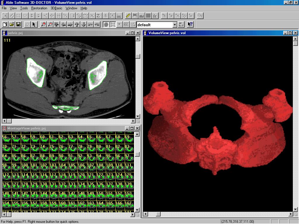
In this new release, 3D-DOCTOR’s volume and surface algorithms have been further optimized to perform volume and surface rendering on a Pentium class PC in real time. The polygon-based surface rendering algorithm creates 3D surface models using contours or object boundaries generated from automatic image segmentation. 3D models can be saved to standard 3D formats, including AutoCAD DXF, 3D Studio, STL, or VRML. The proprietary voxel-based volume rendering supports maximum density, transparency, direct voxel, and ray casting methods and requires much less memory and CPU speed than conventional ray-tracing based algorithms.
In addition, this new release has implemented other high end 3D image processing functions, including 3D image registration, automatic slice alignment, texture-based image segmentation, image mosaic, and improved deconvolution algorithms.
"The 3D imaging market is growing rapidly. With Windows-based imaging software like 3D-DOCTOR, doctors can easily get CT or MRI images to their desktop PC through high speed networks, and perform 3D image rendering and analysis on their PC, or even a laptop computer. The low cost of PC-based software and hardware has made 3D imaging and volume visualization practical and affordable today. It is also possible to use it routinely in a variety of settings, not just in places where mainframe computers or high-end Unix systems are available," said Dr. Y. Ted Wu, founder and president of Able Software.
A free fully functional demo version is available at: http://www.ablesw.com/3d-doctor
World's Leading Research Organizations Choose 3D-DOCTOR
(Aug. 1999) 3D-DOCTOR provides advanced 3D imaging, rendering, and deconvolution functions in a single easy-to-use package running on a PC platform. With 3D-DOCTOR installed, one can immediately have a powerful 3D imaging station on his or her own desktop.
It takes only a few steps to create a 3D model and a 3D rendering from a stack of 2D images. 3D volume calculations can be made from your 3D model. Image measurements can be made with a few mouse clicks. The support to commonly used 3D file formats, such as DXF, STL, VRML and 3DS, makes it easy to bring the 3D data to other systems for visualization and analysis or back into 3D-DOCTOR for display. The 3DBasic scripting tool allows the user to write customized 3D imaging programs and many of the online examples can be used directly.
Because
of its advanced functionality and ease-of-use, 3D-DOCTOR has been
chosen and
is currently being used by leading research
organizations around the world, including MIT (Massachusetts
Institute of Technology), Massachusetts General Hospital (MGH),
Johns Hopkins Univ., SUNY Buffalo, Univ. of Saskatchewan
(Canada), US Navy, US Army, Volkswagen AG (Germany), ABB
Combustion Eng. Sys., Univ. of Pittsburgh, Alberta Research
Council (Alberta, Canada), The Research
Institute of Molecular
Pathology (Austria), Phase 3 Imaging Systems, Monash Univ.(Australia),
American Science & Engineering Inc,
MicroVision (France), Eberhard-Karls-Universitat Tuebingen
(Germany), Minus 9 Technology (Canada), Kanazawa Institute of
Technology (Japan), Univ. of Zilina (Slovakia), Kyung Hee
University (South Korea) and many others.
(Alberta, Canada), The Research
Institute of Molecular
Pathology (Austria), Phase 3 Imaging Systems, Monash Univ.(Australia),
American Science & Engineering Inc,
MicroVision (France), Eberhard-Karls-Universitat Tuebingen
(Germany), Minus 9 Technology (Canada), Kanazawa Institute of
Technology (Japan), Univ. of Zilina (Slovakia), Kyung Hee
University (South Korea) and many others.
(The 3D image on the right is courtesy of G. Martins and Dr. W.J. Sigurdson, Director, Confocal Microscope and 3-D Imaging Facility at SUNY Buffalo.)
3D-DOCTOR Supports DXF, STL, VRML, 3DS, and Raw Triangle Formats
(Aug. 1999) Able Software has released a new version for its 3D imaging, rendering, and deconvolution software, 3D-DOCTOR 2.0. 3D-DOCTOR includes 3D image segmentation, 3D surface and volume rendering, deconvolution, 3D registration, and fusion functions for microscopy imaging and medical imaging applications.
3D-DOCTOR provides tools to define multiple regions of interest for both fully automatic and interactive 3D segmentation to extract object boundaries. Object boundaries can be modified and manipulated using the vector-based editing tools. 3D surface model and volume rendering are then created from the object boundaries. The 3D model data can be exported to AutoCAD DXF, 3D Studio (3DS), STL, VRML, Raw Triangle, and other 3D formats. Import of 3D data files created by other systems have been implemented and it now supports DXF, STL, and Raw Triangle formats.
Supported image formats include DICOM, TIFF, BMP, and other non-standard image formats.
This new release has implemented the 3DBasic scripting language for writing customized program using 3D-DOCTOR's advanced image processing and rendering functions. 3DBasic is a Basic-like scripting language that allows the programming of sophisticated 3D imaging tasks running in both batch and interactive mode.
A fully functioning trial version is available for download at http://www.ablesw.com/3d-doctor. Sample images are available at the http://www.ablesw.com/3d-doctor/images.html.
New International Distributors for 3D-DOCTOR
(July 1999) Able Software has appointed several new international distributors to serve the rapidly expanding international market for its 3D-DOCTOR software, an advanced 3D imaging, rendering, and deconvolution software for the PC platform.
For Japan, Solution Systems, Inc. (Tokyo, Japan, TEL:+81-474-24-6308 FAX:+81-474-24-6327 E-mail : kyorimitsu@msn.com Home Page: http://www.solution-systems.com/) will promote 3D-DOCTOR to the medical imaging and bio-medical imaging market in Japan. In addition, they will provide 3D-DOCTOR training courses to users in the Asia Pacific region.
Paultec (TEL: 82-2-546-5233 FAX: 82-2-546-5242 E-mail: navikim@chollian.net ), a leading imaging software supplier, will be the distributor for South Korea. "We are very happy to become the 3D-DOCTOR distributor in Korea. With the constant technical and marketing support from Able Software, we have had a very successful start introducing 3D-DOCTOR to the Korean market and it has quickly been selected by organizations such as The Department of Mechanical Engineering of Kyung Hee," commented Mr. D.G. Kim, Director of Paultec, Korea.
QBS Software (UK), a leading software distributor in UK, has agreed to sell all Able Software products, including 3D-DOCTOR, to its customers in the United Kingdom.
Daintree Scientific (Tasmania, Australia, TEL: +613-6376.3335 Fax: +613-6376.3334 e-mail: daintree@tassie.net.au), a leading imaging product supplier has become 3D-DOCTOR distributor for Australia. Daintree Scientific has been serving the bio-medical and medical imaging market with years of experience.
New 3DBasic Scripting in 3D-DOCTOR
(May, 1999) Able Software has released a new version for 3D-DOCTOR software, which has implemented the 3DBasic (tm) scripting language for writing customized programs using 3D-DOCTOR's advanced image processing and rendering functions.
3DBasic is a Basic-like scripting language implemented to allow programming with 3D-DOCTOR’s advanced image processing functions to perform sophisticated tasks in batch mode. A 3DBasic program can be created using a text editor, such as Windows NotePad or use 3D-DOCTOR’s 3DBasic/Create command. Use 3DBasic/Run to run an existing 3DBasic program directly.
3D-DOCTOR new version released
(April, 1999) Able software has announced a release for its 3-D imaging software, 3D-DOCTOR version 2.0. 3D-DOCTOR is used for 3D image visualization, volume rendering, and deconvolution applications by researchers, doctors, and engineers.
In this release, image segmentation has been improved to generate object boundaries from image slices and create 3D rendering in a few of steps. 3D contour data can now be imported directly in 3D-DOCTOR for 3D surface rendering and modeling without using an image. 3D surface model can be exported to DXF, 3D Studio, VRML, and other 3D formats.
New algorithms have been developed for 3D image restoration and de-blurring using maximum entropy based deconvolution methods. The 3D surface and volume rendering functions are improved to accommodate user defined scaling factors in all three dimensions. A new automatic image slice alignment function is implemented to correct image misalignment automatically using maximum likelihood image matching technique.
3D-DOCTOR VERSION 2.0 RELEASED
(July, 1998) Able Software Corp. has announced the release of 3D-Doctor version 2.0. 3D-Doctor is an advanced 3D image visualization, rendering, and restoration software used for medical, industrial, microscopy, and scientific 3D imaging applications.
This new version has
implemented a number
of new features and
improvements based on feedback received from users of previous
versions.
Improvements include new intelligent 3D segmentation, fast surface
rendering, improved volume rendering methods, batch rendering,
support of multiple 3D objects, 3D image registration, and image
fusion.
With 3D-Doctor, 3D images in DICOM, TIFF, BMP, or a non-standard format can be opened and visualized in both a 2D plane and a 3D volume and 3D surface view. An irregular region of interest is then defined and used with one of the three segmentation methods to extract object boundaries automatically. Interactive editing and post-processing functions are provided to improve boundary lines obtained from automatic segmentation. A 3D surface is then created using the proprietary 3D surface rendering functions developed by Able Software. Users can choose from either fast simple surface rendering or complex surface rendering. Surface data consists of more than 1 million 3D triangles and is displayed in 3D at any viewing angle or animated along a viewing axis. Volume can also be rendered by combining the image voxels and object boundaries. Batch rendering function allows surface rendering of a large number of images with just a single command.
According to Dr. Yecheng Wu, founder and president of Able Software, "Because of the high performance and low cost, 3D-Doctor software has been warmly received by users doing 3D imaging and rendering applications around the world, including organizations such as United States NAVY, United States ARMY, Volkswagen AG (Germany), Alberta Research Council (Alberta, Canada) and Dept. of Neurology, University of Pittsburgh. Traditionally, people spend up to 40 hours to create a 3D rendering from a raw image. But with 3D-Doctor, this can be reduced to just a few of minutes, thanks to 3D-Doctor's advanced integration of raster and vector system architecture and easy to use interactive editing tools."
3D-DOCTOR HIGHLIGHTED IN THE JUNE ISSUE OF ADVANCED IMAGING
3D-DOCTOR software was highlighted by Advanced Imaging magazine in the article "Image Analysis Software Markets and 3D Modeling: More Than Meets the Eye.", written by Advanced Imaging's contributing editor, Len Yencharis.
In the article, the current market trend for 3D imaging and new technology development has been evaluated. For 3D imaging and rendering software, the author wrote: "Another ambitious effort is 3D-DOCTOR from Able Software, which includes 3D image rendering, volume visualization, image processing and image analysis for CT, MRI, microscopy, ultrasound and industrial testing applications. With 3D-DOCTOR, the object boundaries are extracted using either automatic or interactive 3D image segmentation and used directly for 3D surface and volume rendering. The 3D surface data can be exported as a raster image or vector file for 3D modeling."
3D-DOCTOR is developed by Able Software with years of research and development effort and extensive collaboration with medical doctors and scientists involved in 3D imaging and rendering applications. 3D-DOCTOR was officially released in May 1998, and is currently available for Windows (9X/NT/2000/ME/XP/VISTA) platforms.
3D-DOCTOR SOFTWARE RELEASED
(May, 1998) Able Software Corp. has announced the release of a new software product 3D-DOCTOR. 3D-DOCTOR is developed for 3D image rendering, volume visualization, image processing, and analysis used by CT, MRI, microscopy, ultrasonic, and industrial testing applications.
3D-DOCTOR provides advanced tools to visualize 3D images and volumetric data (CT, MRI, Microscopy, and other image types) stored in DICOM, TIFF, BMP, or other formats.
Object boundaries are extracted using either fully automatic or interactive 3D image segmentation and used directly for 3D surface and volume rendering. A 3D rendering is constructed from 2D image slices in a few seconds. 3D surface data can be exported either as a raster image or a vector file (DXF) with triangular faces for 3D modeling and other applications.
A 3D image object can be split into smaller sections and viewed from any direction. Image measurements, including length, area, surface area, volume, image profile, and image histograms, are obtained easily by drawing interactively with the mouse.
3D image restoration is supported by deconvolution using either nearest neighbor or maximum entropy methods. The restoration can remove or reduce the effect of image blurring or degradation introduced in the imaging process when it can be modeled with a point spread function (PSF).
3D image registration allows a 3D image to be geometrically corrected and registered to another image and can further create a fusion image by combining the two images together. Image fusion makes it possible to see certain image features which may otherwise be invisible or not easily seen if not combined with images acquired by other means.