3D IMAGING AND MODELING USING CT, MRI AND OTHER IMAGES
Click here to watch a 3D-DOCTOR demo video
Input Image: CT, MRI, PET and other cross-sectional images in DICOM, TIFF, BMP, JPEG, Interfile, PNG, PGM, GIF, Raw Image Data, and other uncompressed image formats. Image files in various vendor specific formats can be easily read using 3D-DOCTOR's universal image configuration and input function. Both grayscale (8-bit and 16-bit) and color images are supported. Scanned CT/MRI films can easily be cropped using the template-based function for 3D imaging applications.
|
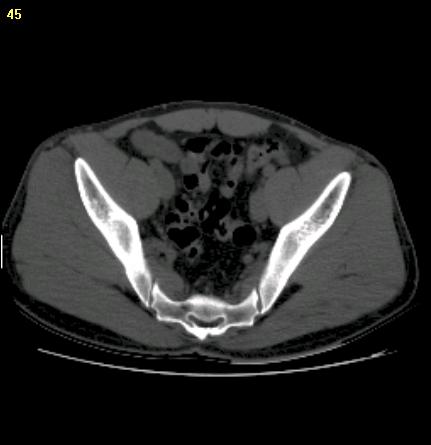
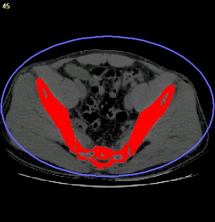
Pelvis CT Image |
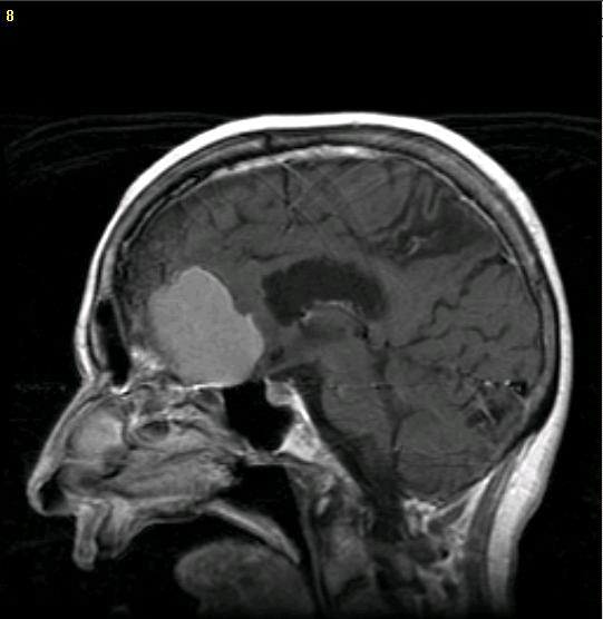
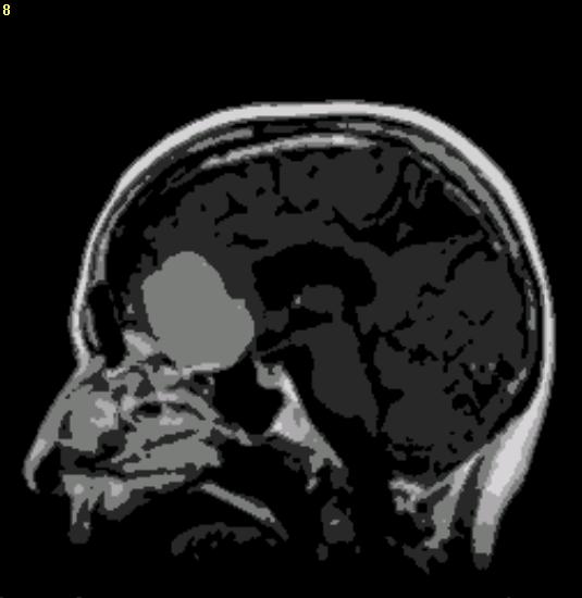
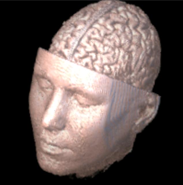
Head MR Image with Brain Tumor |
Segmentation Tools: the fully automatic texture-based segmentation for grayscale and color images, the thresholding-based Interactive Segmentation for CT images, the region-based Object Segmentation and the easy-to-use polygon-based manual tracing.
Threshold-based Interactive Segmentation |
Texture-based fully automatic segmentation |
3D Model Export: STL (ASCII and Binary), AutoCAD DXF, 3D Studio (3DS), IGES, VRML, Wavefront OBJ, PLY, raw triangles, and other 3D graphics file formats for rapid prototyping, 3D printing, finite element analysis, animation and visualization applications. Click here to download 3D sample files to check the format.
Once object boundaries are generated from segmentation, 3D mesh models are created in using one of the surface rendering functions. Unlimited number of objects representing different tissues are supported .
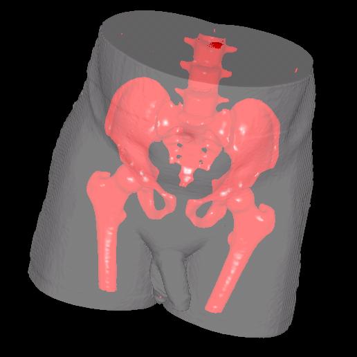
3D mesh model created from CT image |
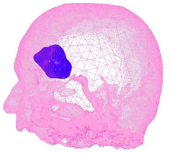
3D mesh model of tumor and skin |
Measurement and Quantitative Analysis
Measurements include area, 3D surface area, 3D volume, distance, profile, and image region histogram. Quantitative report of objects (number of pixels, pixel density, statistical data) is easily obtained for research and analysis.
3D Visualization
Real-time 3D surface rendering and volume rendering for visualization applications. 3D Animation. Create 3D models from CT, MRI and microscopy images and use 3D-DOCTOR's advanced animation control function to move each object independently and generate an AVI movie file. Click here to see an example.
3D volume rendering from MRI image |
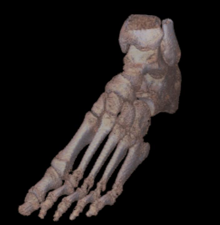
3D volume rendering of a foot |
Registration and Fusion
Images of different modalities
can be registered using the 3D image registration function and fused
together to create a new fusion image. The Reslice command takes an
image with variable spacing between slices to create a new image of
evenly space slices or simply create a new image with a different slice
thickness. An image can be resliced along a user-defined axis so
more accurate measurements can be made for certain features.
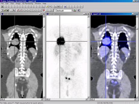
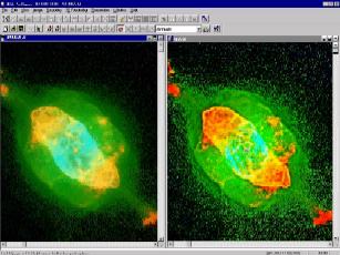
Fusion of a CT and PET image |
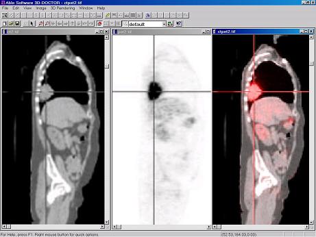
Fusion of a CT and PET image |
Advanced 3D Image Processing
3D Computed Tomography: Create parallel cross-section, volume images using x-ray images taken at angles around an object. Turn your x-ray machine into a full CT system using 3D-DOCTOR.
Other image processing functions include: template-based scanned film cropping, volume resizing, 3D image filtering, Image rotation, orientation adjustment, contrast adjustment, background removal, image combination, linear feature extraction, pattern recognition, segmentation, image mosaic, and color classification can all be performed on your 3D images.
3D Image Restoration by Deconvolution: 3D-DOCTOR provides two highly efficient deconvolution methods for 3D image restoration and reconstruction, a fast nearest neighbor algorithm and an iterative maximum entropy algorithm.
3DBasic scripting language will let you create your own Basic-like sophisticated programs using 3D-DOCTOR's advanced imaging and rendering functions quickly.
Deconvolution of an image
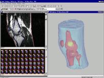
from Hubble Space Telescope 3D models created from a MRI
knee image